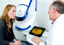
Why optomap ultra-widefield retinal imaging?

Your retina (located in the back of your eye) is the only place in the body where blood vessels can be seen directly. This means that in addition to eye conditions, signs of other diseases (for example, stroke, heart disease, hypertension, and diabetes) can also be seen in the retina. Early signs of these conditions can show on your retina long before you notice any changes to your vision or feel pain. While eye exams generally include a look at the front of the eye to evaluate health and prescription changes, a thorough screening of the retina is critical to verify that your eye is healthy.
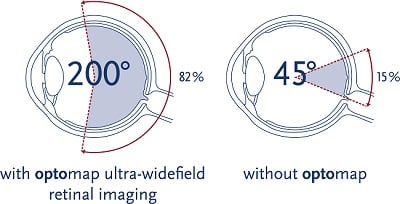
Early detection
is essential to reduce the risk to your sight and health

What is an optomap Image?

Getting an optomap image is fast, painless, and comfortable. Nothing touches your eye at any time. It is suitable for the whole family. To have the exam, you simply look into the device one eye at a time (like looking through a keyhole) and you will see a flash of light to let you know the image of your retina has been taken.

Under normal circumstances, dilation drops might not be necessary, but your eye care practitioner will decide if your pupils need to be dilated depending on the health of your eyes. The image capture takes less than a half-second and they are available immediately for you to see your own retina. You see exactly what your eye care practitioner sees - even in a 3D animation.

Early signs of disease can be present in the periphery of your retina and remain undetected for a long time when using traditional methods. The optomap ultra-widefield retinal image is a unique technology that captures more than 80% of your retina in a single image while traditional imaging methods typically only show 15% of your retina at one time.


Benefits of an optomap Image

The benefits of having an optomap ultra-widefield retinal image taken are:

- optomap facilitates early protection from vision impairment or blindness

- Early detection of life-threatening diseases like cancer, stroke, and cardiovascular disease

The unique optomap ultra-widefield view helps your eye care practitioner detect early signs of retinal disease more effectively and efficiently than with traditional eye exams.

Early detection means successful treatments can be administered and reduces the risk to your sight and health.


Questions about optomap...

Frequently Asked Questions about an optomap

Why is a retinal exam so important?

Some of the first signs of diseases such as stroke, diabetes and even some cancers can be seen in your retina, often before you have other symptoms.  An optomap makes it easier to see them.

Is an optomap safe for children?

Yes.  In fact, many vision problems begin in early childhood, so it's important for children to receive quality routine eye care.

What is an optomap?

The optomap is a digital image of the retina produced by Optos scanning laser technology.  It is the only technology that can capture 82% view of your retina at one time.

Does it hurt?

No.  It is completely comfortable and the scan takes less than a second.

How will optomap benefit me?

The ultra-widefield optomap may help your eye doctor detect problems more quickly and easily.  Unlike traditional retinal exams, the optomap image can be saved for future comparisons.

How often should I have an optomap?

This is a decision that should be made by your doctor.  However, it is generally recommended that you have an optomap each time you have an eye exam.

Are there side effects?

optomap images are created by non-invasive, low-intensity scanning lasers.  No adverse health effects have been reported in over 150 million sessions.