Patient Stories

Learn how optomap has helped eye care professionals save sight and even save lives.


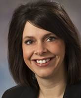
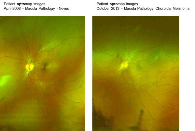
Denise Kniefel, OD knows the value of the optomap exam because she has been a customer of Optos since 2004. No stranger to the technology, Dr. Kniefel started imaging her retina as a matter of course. In 2008 at a tradeshow, she stopped in to talk to the representatives from the company. Optos had recently unveiled new technology that allowed for even better optomap imaging than in the past. Dr. Kniefel sat down and had her retinal image captured. A nevus was detected that she hadn’t previously seen.

The image prompted Dr. Kniefel to see her ophthalmologist and have a fluorescein angiography and an OCT exam done. She did indeed have a nevus, which is essentially a freckle in the eye, and needs to be routinely monitored. Just like a freckle on your skin, it can change and become cancerous. Her doctor told her if the nevus did change into cancerous melanoma of eye, she would notice changes to her vision.

Denise continued to image herself every six months using the optomap, and in 2013, she noticed that the nevus looked different but she was not experiencing any vision loss. She went back to her retinal specialist who told her the nevus was now presenting as a very small choroidal melanoma. Dr. Kniefel went to Evangelos Gragoudas, MD at Massachusetts Eye and Ear Infirmary in December of 2013 for proton beam radiation. Because it is so soon after her treatment, her prognosis is unclear. However, because of the optomap technology, the cancer was caught at an extremely early stage, the best case scenario for a positive outcome. 

Only 6 out of a million people are diagnosed with choroidal melanoma (cancer of the eye) and most of those go undetected until they are large. Due to her own experience, she is a strong supporter of the optomap technology and recommends it to all of her patients and within her own personal community. She is convinced that the optomap exams that she was using to track changes in her nevus enabled her to get world-class treatment for her melanoma before it could have been detected by a typical dilation fundus exam.

View optomap images


 optomap images

Nakivia's Story

In 2009, Nakivia visited Dr. Andrew Kraft's office in Enid, OK. During her visit she received information about optomap . Despite the process not being covered by her insurance provider, she agreed to her first screening to establish a baseline. In May of 2013, she decided to have another optomap done to see if there had been any changes in her eye pathology. The comparison between her 2013 results and previous screenings showed that her optic nerve was bending and there was hemorrhaging of her blood vessels. Within 4 hours of receiving her optomap at what was supposed to be a routine exam, she saw Dr. Barry Pollard, and received a specialized MRI.

The results of this MRI showed that Nakivia had a substantial brain tumor that was causing the optic nerve damage and hemorrhaging. Nakivia received her surgery and eight months later, she is not only alive but her vision has improved by more than 50% without corrective lenses. Nakivia's doctors suggested that without the immediate attention based on the results from the optomap exam, she would have most likely had a stroke or seizure within a few months.

View optomap images

Nakivia's Story

Nakivia's Story optomap images

Tori's Story

At the age of 8, Tori and her mom visited Dr. Scott Thiele, Jacksonville, FL, for her first eye exam which included an optomap. She had a slight blur in vision, but no other symptoms. The optomap revealed a large retinal detachment, which occurs when the retina lifts or detaches from its normal position. Based on the size of the detachment, Dr. Thiele suspected cancer and immediately referred her to the hospital. She was diagnosed with a rare, large choroidal melanoma and unfortunately her eye had to be removed. Tori's mother credits the optomap for discovering the tumor that could have been fatal if left undetected.

Tori's Story

Cainan's Story

In preparation for Back to School, the mother of a 6-year-old boy, Cainan, scheduled an eye exam. Cainan received an optomap the previous year and Dr. Jacqueline Campisi, Mystic, CT, compared his optomap images and noticed that the optic nerves, which transmit visual messages from the retina to the brain, in both eyes were swollen. Cainan was referred to a pediatric ophthalmologist and pediatric neurologist and was later diagnosed with papillitis aseptic meningitis due to Lyme disease. Lyme disease is an illness caused by a bacteria transmitted through the bite of infected ticks. Cainan was hospitalized at the time, but was released after a few days and is now healthy.

Cainan's Story