Retinal Detachment
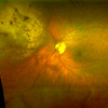
When the retina detaches, it is lifted or pulled from its normal position. If not promptly treated, a retinal detachment can cause permanent vision loss. Anyone can get a retinal detachment; however, they are far more common in nearsighted people, those over 50, those who have had significant eye injuries, and those with a family history of retinal detachments.
What are the different types of retinal detachments?
There are three different types of retinal detachments:
-
Rhegmatogenous [reg-ma-TAH-jenous] – A tear or break in the retina causes it to separate from the retinal pigment epithelium (RPE), the pigmented cell layer that nourishes the retina, and fill with fluid. These types of retinal detachments are the most common.
-
Tractional – In this type of detachment, scar tissue on the retina's surface contracts and causes it to separate from the RPE. This type of detachment is less common.
-
Exudative – Frequently caused by retinal diseases, including inflammatory disorders and injury/trauma to the eye. In this type, fluid leaks into the area underneath the retina (subretina).
Text, images and photographs taken from National Eye Institute (NEI), National Institutes of Health (NIH). For more information on retinal detachment visit www.nei.nih.gov.
Who is at risk for a retinal detachment?
Although anyone can experience a retinal detachment, people with certain eye conditions are at increased risk. Some examples of these conditions include posterior vitreous detachment, lattice degeneration, x-linked retinoschisis, degenerative myopia, and uveitis. Injuries to the eye or head can also cause a retinal detachment.
What are the symptoms of a retinal detachment?
Symptoms include a sudden or gradual increase in the number of floaters and/or light flashes in the eye or the appearance of a curtain over the field of vision. A retinal detachment is a medical emergency. Anyone experiencing the symptoms of a retinal detachment should see an eye care professional immediately.
How is a retinal detachment treated?
Small holes and tears are treated with laser surgery or a freeze treatment called cryopexy. These procedures are usually performed in the doctor's office. During laser surgery, tiny burns are made around the hole to "weld" the retina back to into place. Cryopexy is a similar procedure that freezes the area around the hole.
Retinal detachments are treated with surgery that may require the patient to stay in the hospital. In some cases a scleral buckle, a tiny synthetic band, is attached to the outside of the eyeball to gently push the wall of the eye against the detached retina. If necessary, a vitrectomy may also be performed to treat more severe cases. During a vitrectomy, the doctor makes a tiny incision in the sclera (white of the eye). Next, a small instrument is placed into the eye to remove the vitreous. Salt solution is then injected to into the eye to replace the vitreous.
Early treatment can usually improve the vision of most patients with retinal detachments. Some patients, however, will need more than one procedure to repair the damage.
An eye care professional who has examined the patient's eyes and is familiar with his or her medical history is the best person to answer specific questions.